EPMA-8050G - Aplicações
Electron Probe Microanalyzer
Ultra High Resolution Mapping
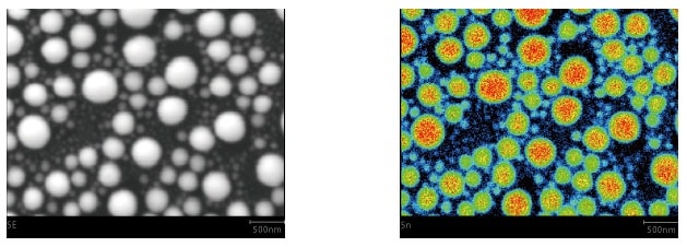
A mapping analysis of Sn balls on carbon was performed at a magnification of 30,000x. Even Sn particles a mere 50 nm in diameter, visible in the SE image (left), can be confirmed precisely in the X-ray image (right).
Ultra High Sensitivity Mapping
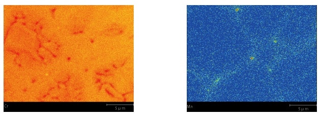
A mapping analysis of stainless steel was performed with a beam current of 1 μA at a magnification of 5,000x. (Left) The distribution of phases with slightly different Cr concentrations is precisely captured. (Right) The system succeeds in visualizing a distribution of Mn content under 0.1 %.
Distribution of Ag and Cu in Lead-Free Solder
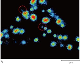
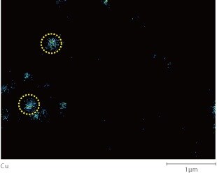
This data is from a mapping analysis of areas in lead-free solder containing a large amount of Ag. (Accelerating voltage: 10 kV; beam current: 20 nA) The shape of the particles in the Ag X-ray image agrees well with the shape of the particles in the BSE image (COMPO). It is evident that the particles with a diameter of about 0.1 μm, indicated by the red dashed lines, are also Ag particles. In addition, the existence of Cu-containing particles can also be confirmed, as shown by the yellow dashed lines.

Metallic Elements in Biological Tissues (DDS)
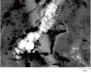
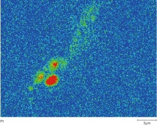
This EPMA element imaging data shows the existence of platinum (Pt), a component (platinum complex) of the anticancer drug carboplatin, delivered (by a DDS) into the tissue of a head and neck tumor in a mouse. By binding with DNA strands, the genetic substance inside the cancer cells, the carboplatin prevents cell division (DNA replication), destroying the cells. The element imaging clearly shows the manner in which anticancer drug is delivered within the cancer cells.
